Leukocyte Count Correlates with Platelet Count in Pediatric Typhoid Fever
DOI:
https://doi.org/10.58184/miki.v4i2.938Keywords:
typhoid fever, leukocyte count, platelet count, pediatric typhoid fever, childrenAbstract
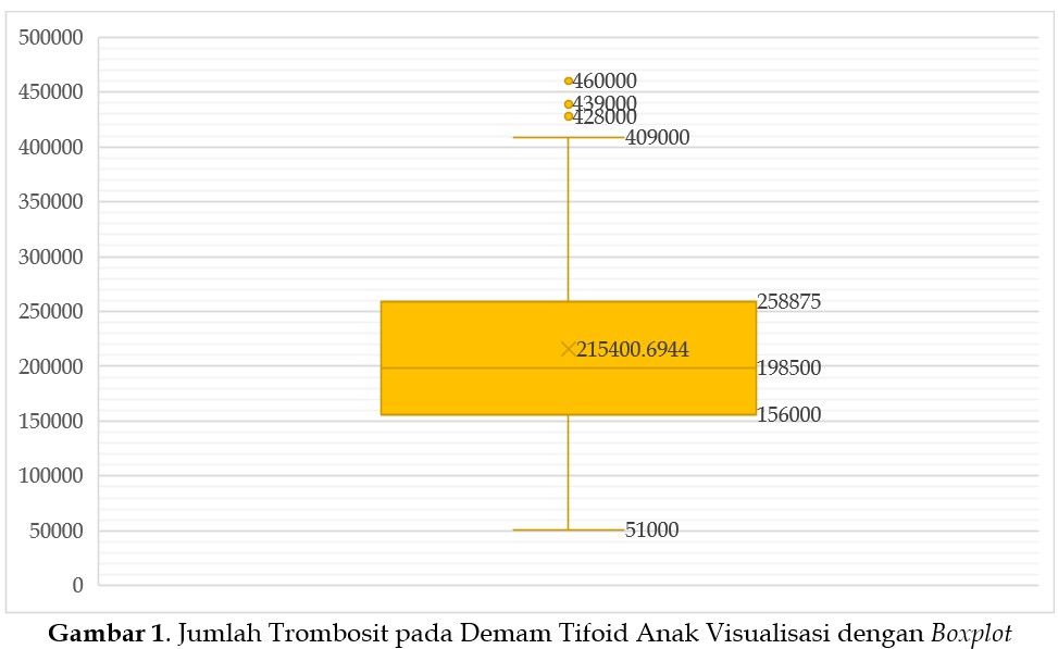
Typhoid fever in children remains a health problem in developing countries, with hematological changes such as leukocyte and platelet abnormalities being frequently observed. This study aimed to analyze the relationship between leukocyte and platelet counts in pediatric patients with typhoid fever. A cross-sectional design was employed using secondary data from the Clinical Pathology Laboratory of RSDI Banjarbaru City. A total of 72 children aged 5–13 years who met the inclusion criteria (diagnosis of typhoid fever by Tubex TF, no prior antibiotic therapy) were analyzed. Leukocyte and platelet measurements were performed using a hematology analyzer, and data were analyzed using simple linear regression (SPSS version 24). Results showed a mean age of 8.2 years, with a higher proportion of males (59.7%). Leukocyte distribution was highly variable, ranging from severe leukopenia (1200–1248/mm³) to extremely high values. Platelet distribution was also wide, with very low values of 51,000/mm³ and high outliers up to 460,000/mm³. Regression analysis yielded the equation y = 13.74x + 118195 with a coefficient of determination (R²) of 0.4998, indicating that approximately 50% of the variability in platelet count could be explained by leukocyte count. Pathophysiologically, this relationship is mediated by proinflammatory cytokines (TNF-α, IL-1, IL-6) and leukocyte-platelet aggregation induced by Salmonella typhi endotoxin. In conclusion, there is a moderate positive correlation between leukocyte and platelet counts in children with typhoid fever. These hematological parameters have the potential to serve as adjunctive diagnostic tools and for risk stratification, particularly in settings with limited blood culture facilities.
Downloads
References
Ahmed, S., Ali, F., & Areej, H. A. (2023). 362 Assessing differences in hematologic changes caused by XDR and non-XDR strains of salmonella typhi in pediatric patients with typhoid fever. British Association of General Paediatrics, A111.2-A111. https://doi.org/10.1136/archdischild-2023-rcpch.186
Aird, W. C. (2003). The role of the endothelium in severe sepsis and multiple organ dysfunction syndrome. Blood, 101(10), 3765–3777. https://doi.org/10.1182/blood-2002-06-1887
Akbayram, S., Parlak, M., Dogan, M., Karasin, G., Akbayram, H., & Karaman, K. (2016). Clinical and Hematological Manifestations of Typhoid Fever in Children in Eastern Turkey. West Indian Medical Journal. https://doi.org/10.7727/wimj.2014.354
Chaudhry, A. R., Kazi, M. Y., Usman, M., & Ayub, R. (2022). Frequency of leukocytosis in culture proven enteric fever in children. The Professional Medical Journal, 29(06), 823–828. https://doi.org/10.29309/TPMJ/2022.29.06.6647
Dhillon, S. P. S., Neeraj, L., Sehajpreet, S., Vijay, G., Tanushree, J., & Narinder, S. (2017). To study haematological profile of Enteric fever patients. Int J Curr Res Med Sci, 3(7), 24–29. https://doi.org/10.22192/ijcrms.2017.03.07.004
Hidayatallah, Z. H. (2024). Comparative analysis of azithromycin and cefixime in the treatment of typhoid fever. Journal of Advanced Zoology, 45, 48–56. https://doi.org/10.53555/jaz.v45iS2.3756
Khan, A., Khan, I., Babar, A. N., Khan, Y., Shah, G., & Khan, M. I. (2024). Effectiveness of Oral Azithromycin in Treating Enteric Fever: A Hospital-Based Study on Pediatric Patients. Cureus, 16(8), e67024. https://doi.org/10.7759/cureus.67024
Kuncahyono, G. H., & Airlangga, E. (2025). Correlation of Leukocyte and Lymphocyte Counts With Positive Tubex Scores in Pediatric Typhoid Fever Patients. AVERROUS: Jurnal Kedokteran Dan Kesehatan Malikussaleh, 1–15. https://doi.org/10.29103/averrous.v11i2.24788
Lestari, A. A. W., Sukrama, I. D. M., & Nurmansyah, D. (2019). The earthworm (Lumbricus rubellus) extract decreased amino transaminase enzyme level and number of bacterial colony in male wistar rats infected with Salmonella typhimurium. Biomedical and Pharmacology Journal, 12(1), 325–332. https://doi.org/10.13005/bpj/1643
Malik, A. S. (2002). Complications of Bacteriologically Confirmed Typhoid Fever in Children. Journal of Tropical Pediatrics, 48(2), 102–108. https://doi.org/10.1093/tropej/48.2.102
Mushtaq, S., Bhat, A. A., Rather, G. N., Akhter, R., Bhat, I., & Wani, T. (2017). Clinical profile of enteric fever in tertiary care hospital of Kashmir. International Journal of Contemporary Pediatrics, 4(5), 1754. https://doi.org/10.18203/2349-3291.ijcp20173779
Ndako, J. A., Olisa, J. A., Ifeanyichukwu, I. C., Okolie, C. E., Ojo, S. K. S., & Jegede, S. L. (2020). Predictive evaluation of pediatric patients based on their typhoid fever status using linear discriminant model. Medical Hypotheses, 144, 110264. https://doi.org/10.1016/j.mehy.2020.110264
Nurmansyah, D., Fayumi, S., Shalehatun Nisa, P., Sasmitha, M., & Arsyad, M. M. M. (2025). Lymphocyte Cell Count Profile based on Widal Titer On Pediatric Typhoid Fever. Jurnal Analis Medika Biosains (JAMBS), 12(2), 102-107. https://doi.org/10.32807/jambs.v12i2.480
Nurmansyah, D., Nisa, S., Puspawati, P., Fayumi, S., Mudzakkir, M., & Sasmitha, M. H. (2025). Profil Jumlah Leukosit Berdasarkan Nilai Titer Widal pada Kasus Demam Tifoid Anak. JSN : Jurnal Sains Natural, 3(2), 107–111. https://doi.org/10.35746/jsn.v3i2.845
Paradise, G., Agraini, A., & Fitriana, E. (2024). Study of Platelocyte Count on Patient Fever Typhoid at Baiturrahim Jambi Hospital. Proceeding International Conference Health Polytechnic of Jambi, 3, 17–21. https://doi.org/10.35910/icohpj.v3i0.822
Patel, D., Patel, D., & Virpariya, M. (2024). Evaluation of neutrophil–lymphocyte ratio and platelet–lymphocyte ratio in children with typhoid fever. Int J Acad Med Pharm, 6(3), 38–44. https://doi.org/10.47009/jamp.2024.6.3.9
Rafiq, F., Shafqat, F., Kahif, Z., Rehman, M. U., Ali, S. S., & Warriach, S. Z. (2022). Histopathological and Haematological Manifestations of Typhoid Fever in Pediatric Age Group. Pakistan Journal of Medical and Health Sciences, 16(3), 60–62. https://doi.org/10.53350/pjmhs2216360
Ringoringo, H. P., Wahyuni, I. I., Panghiyangani, R., Hartoyo, E., & Lao, R. (2021). Hematological profile of children under five years with typhoid fever at Idaman Banjarbaru Hospital, Indonesia. Bali Medical Journal, 11(2), 775–778. https://doi.org/10.15562/bmj.v11i2.2669
Samatra, D. P. G. P., Mahadewa Tjokorda, G. B., Sukrama, D. M., Dewi, N. W. S., Praja, R. K., Nurmansyah, D., & Widyadharma, I. P. E. (2017). Extract of earthworms (Lumbricus rubellus) reduced malondialdehyde and 8-hydroxy-deoxyguanosine level in male wistar rats infected by salmonella typhi. Biomedical and Pharmacology Journal, 10(4), 1765–1771. https://doi.org/10.13005/bpj/1290
Semple, J. W., Italiano, J. E., & Freedman, J. (2011). Platelets and the immune continuum. Nature Reviews Immunology, 11(4), 264–274. https://doi.org/10.1038/nri2956
Sethuraman, N., Priyadharshini, D., Clinton, M., Sridhar, M., & Krishna, V. (2024). Clinical Profile and Prescription Patterns in Culture-proven Enteric Fever in Children. Pediatric Infectious Disease, 6(1), 6–9. https://doi.org/10.5005/jp-journals-10081-1408
Sharma, P. K., Vinayak, N., Aggarwal, G. K., Srivastava, R. D., & Aggarwal, P. K. (2021). Clinical Profile, Laboratory Findings, Antimicrobial Resistance and Antibiotic Usage in Children with Culture Positive Enteric Fever. The Indian Journal of Pediatrics, 88(2), 180–181. https://doi.org/10.1007/s12098-020-03415-8
Tahir, N., Khattak, S. A. K., Jamal, N., Shaikh, G. M., Ahmad, R., & Ahmed, W. (2024). Haematological manifestations in children with extended drug-resistant Salmonella typhi infections in a tertiary care hospital. Journal of Liaquat University of Medical & Health Sciences, 23(01), 53–57. https://doi.org/10.22442/jlumhs.2024.01082
Tobing, J. F. (2024). Demam tifoid. Ikra-Ith Humaniora: Jurnal Sosial dan Humaniora, 8(2), 463–470.
Downloads
Published
How to Cite
Issue
Section
License
Copyright (c) 2026 Dian Nurmansyah, Lala Foresta Valentine Gunasari, Puspawati, Maya Herliana Sasmitha

This work is licensed under a Creative Commons Attribution-ShareAlike 4.0 International License.