Effect of Film Focus Distance Variation on Anode Heel Effect and Radiographic Image Quality Based on Pixel Value Parameters
DOI:
https://doi.org/10.58184/miki.v4i2.978Keywords:
anode heel effect, FFD, pixel value, radiographyAbstract
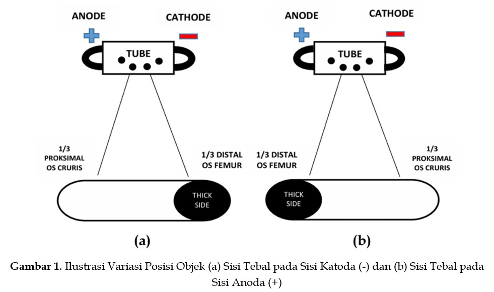
This study aimed to analyze the effect of variations in focus-to-film distance (FFD) on the anode heel effect and radiographic image quality, based on the Pixel Value (PV) parameter. A quantitative experimental approach was employed. The testing was conducted using a phantom articulatio genu with FFD variations of 90 cm, 100 cm, and 120 cm, and object positions varied between the cathode and anode sides. The results indicated that the anode heel effect was more effectively utilized at shorter FFD when the thick side of the object was positioned on the cathode side. The highest PV value was obtained under this condition, whereas a decrease of approximately 11% was observed when the thick side was positioned on the anode side. Furthermore, an FFD of 100 cm produced lower PV standard deviation (PVSD) values, resulting in more homogeneous images with minimal noise. These findings demonstrate that variations in FFD and object positioning influence the distribution of X-ray intensity and radiographic image quality. Appropriate selection of FFD and placement of the thick side of the object on the cathode side can produce more uniform radiographs in terms of sharpness, brightness, and density.
Downloads
References
Adi, E. P., & Iqbal, M. (2020). Comparison of radiograph image information on lumbar vertebrae examination using the application of the anode heel effect theory. Journal of Physics: Conference Series, 1517(1), Article 012052. https://doi.org/10.1088/1742-6596/1517/1/012052
Ani, J., Lumanauw, B., & Tampenawas, J. L. A. (2021). The Influence of Brand Image, Promotion, and Service Quality on Consumer Purchasing Decisions on Tokopedia E-Commerce in Manado City. Jurnal Emba, 9(2), 663–674. https://doi.org/10.35794/emba.v10i1.38279
Barati, B., Kaydani, M., Birgani, F. F, Zabihzadeh, M., Tahmasebi, M. J., & Chegeni, N. (2024). The effect of anode, hot and cold cathode structures used in miniature X-ray tubes on X-ray output using Monte Carlo simulation. In A. Abou Jaoudé (Ed.), Recent advances in Monte Carlo methods. IntechOpen. https://doi.org/10.5772/intechopen.1001904
Chou, M. C. (2021). Evaluation of non-uniform image quality caused by anode heel effect in digital radiography using mutual information. Entropy, 23(5), 525. https://doi.org/10.3390/e23050525
Edirisinghe, S., De-Silva, D., Dissanayake, H., Yasawardene, S., Devmini, M., Pathmaperuma, S., & De-Zoysa, N. (2024). Anatomical Diversity in Femur bones: Understanding the Morphological Variability for Surgical and Prosthetic Applications. International Journal of morphology, 42(1), 162–165. https://doi.org/10.4067/S0717-95022024000100162
Febriansyah, T. N., Pratiwi, B. N., Pramono, S., Sardjono, Y., Triatmoko, I. M., Wijaya, G. S., Prasetio, H., Hidayati, N. R., Nuraeni, N., Ulya, S., & Ismail, Z. (2025). Dose Analysis of Prostate Cancer Therapy with X-Ray Therapy using PHITS Program Version 3.341. Jurnal Teknologi Reaktor Nuklir Tri Dasa Mega, 27(1), 49–58. https://doi.org/10.55981.tdm.2025.7158
Fung, K. K., & Gilboy, W. B. (2000). "Anode heel effect" on patient dose in lumbar spine radiography. The British Journal of Radiology, 73(869), 531–536. https://doi.org/10.1259/bjr.73.869.10884750
Grammens, J., Van Haver, A., Danckaers, F., Booth, B., Sijbers, J., & Verdonk, P. (2021). Small medial femoral condyle morphotype is associated with medial compartment degeneration and distinct morphological characteristics: a comparative pilot study. Knee Surgery, Sports Traumatology, Arthroscopy, 29(6), 1777–1789. https://doi.org/10.1007/s00167-020-06218-8
Kusk, M. W., Jensen, J. M., Gram, E. H., Nielsen, J., & Precht, H. (2021). Anode heel effect: Does it impact image quality in digital radiography? A systematic literature review. Radiography, 27(3), 976–981. https://doi.org/10.1016/j.radi.2021.02.014
Marvellini, R. Y. (2022). Medical Students’ Knowledge Level about X-Ray as a Diagnostic Support Tool at Universitas Kristen Indonesia, Jakarta Batch 2016. International Journal of Health Sciences and Research, 12(2), 318–328. https://doi.org/10.52403/ijhsr.20220242
Prabhu, S., Naveen, D. K., Bangera, S., & Subrahmanya Bhat, B. (2020). Production of x-rays using x-ray tube. In Journal of Physics: Conference Series,1712(1), Article 012036. https://doi.org/10.1088/1742-6596/1712/1/012036
Precht, H., Hansson, J., Outzen, C., Hogg, P., & Tingberg, A. (2019). Radiographers’ perspectives’ on Visual Grading Analysis as a scientific method to evaluate image quality. Radiography, 25, S14–S18. https://doi.org/10.1016/j.radi.2019.06.006
Rayan, A. M., Adam, A., Al-Arabi, G., & Ahmed, M. R. (2025). The applications of X-ray technology in medical imaging: advances, challenges, and future perspectives (A review). Journal of Sustainable Food, Water, Energy and Environment, 1(2), 39–61. https://doi.org/10.21608/jsfw.2025.409882.1003
Rosidah, S., & Felayani, F. (2022). Effect of Heel Effect Anode on Image Homogeneity Based on X-Ray Collimation Light Beam Area. Jurnal EduHealth, 13(01), 400–406.
Rubia-Bullen, I. R. F., Escarpinati, M. C., Schiabel, H., Vieira, M. A. D. C., Rubira, C. M. F., & Lauris, J. R. P. (2007). Digitizing radiographic films: a simple way to evaluate indirect digital images. Journal of Applied Oral Science, 15(1), 14–17. https://doi.org/10.1590/S1678-77572007000100004
Salleh, H., Samat, S., Matori, M. K., Md Isa, M. J., Arshad, M. R., Azizan, S. A., Abdul Rahman, M. F., & Md Zin, M. K. (2014, October 14–16). Heel effect: Dose mapping and profiling for mobile C-arm fluoroscopy unit Toshiba SXT-1000A [Oral presentation]. Research and Development Seminar 2014, Bangi, Malaysia. International Atomic Energy Agency. https://doi.org/10.13140/2.1.3222.0161
Sari, A. R., Al Husnawati, H., Suryono, J., Marzuki, M., & Mulyapradana, A. (2025). Metode Penelitian Kualitatif, Kuantitatif, dan R&D. YPAD Penerbit; Yogyakarta.
Seibert, J. A. (2004). X-ray imaging physics for nuclear medicine technologists. Part 1: Basic principles of x-ray production. Journal of nuclear medicine technology, 32(3), 139–147.
Vicchietti, M.L., Ramos, F.M. & Campanharo, A.S.L.O. Pattern and structural detection in grayscale images through the application of quantile graphs in higher-dimensional spaces. Scientific Reports, 15(1), 44356. https://doi.org/10.1038/s41598-025-27825-w
Downloads
Published
How to Cite
Issue
Section
License
Copyright (c) 2026 Sitti Normawati, Syahrir, Nur Fitri Qalbina Syahdan, Jumardin

This work is licensed under a Creative Commons Attribution-ShareAlike 4.0 International License.